Back to the on-screen lesson ·
d-d absorption, the octahedral splitting from the wavelength of a band, complementary colors, selection rules, and Jørgensen's rule for predicting a splitting.
Paper packet. Every task here also exists on screen, where it is checked automatically; answers written on paper are not assessed by Nydus. When you are back at a device, enter your answers there.
By the end of this lesson you will be able to find $\Delta_o$ from the wavelength of a d-d band, turn it into a stabilization energy, and predict the color of a complex.
Lesson 10 introduced the splitting $\Delta_o$ and the conversion from wavenumbers to kJ/mol, and lesson 11 ranked ligands in the spectrochemical series. Lesson 1 said that transition-metal ions are colored because of their d electrons. From physics you know that light of wavelength $\lambda$ comes in photons of energy $E = hc/\lambda$, so shorter wavelengths carry more energy. This lesson connects all of these.
| Term | What it means |
|---|---|
| d-d transition | The jump of an electron from a lower d orbital to a higher one when a photon is absorbed. |
| Absorption band | The range of wavelengths a complex absorbs, broad for a d-d transition. |
| $\lambda_{\max}$ | The wavelength at the top of an absorption band. |
| Wavenumber, $\tilde\nu$ | The reciprocal of the wavelength, in cm$^{-1}$; it is proportional to energy. |
| Complementary color | The color seen when a band of other colors is removed from white light. |
| Selection rule | A rule saying which transitions absorb strongly and which only weakly. |
| Charge-transfer band | An intense band in which an electron moves between the ligand and the metal. |
An electron in a lower d orbital can absorb a photon and jump to a higher one, but only if the photon's energy matches the gap. For a first-row complex the gap is tens of thousands of wavenumbers, which is the energy of visible light. So the complex absorbs part of the visible spectrum, and we see the rest.
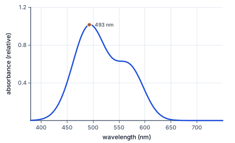
The simplest case is a $d^1$ ion such as $\mathrm{[Ti(H_2O)_6]^{3+}}$. Its one electron sits in $t_{2g}$, and a photon of energy $\Delta_o$ lifts it to $e_g$. The absorption band peaks at the wavelength whose photons carry exactly that energy.
The band peaks at $493$ nm, in the blue-green. To turn a wavelength into a splitting, use wavenumbers, which are proportional to energy. With $\lambda$ in nanometers,
$$\tilde\nu\,(\text{cm}^{-1}) = \frac{10^7}{\lambda\,(\text{nm})},$$
because ten million nanometers make one centimeter. So $\Delta_o = 10^7/493 \approx 20{,}300$ cm$^{-1}$, and with lesson 10's conversion, about $243$ kJ/mol.
The color we see is what is left. The titanium solution absorbs strongly from blue-green to yellow and transmits the red and violet ends of the spectrum, which together look purple. As a rough guide, the color seen is the complement of the color absorbed: absorb violet and the sample looks yellow-green; absorb blue, orange-yellow; absorb green, red-purple; absorb yellow, violet; absorb orange, blue; absorb red, green.
For most ions more than one band appears, because a d electron can be excited in several ways that interact with each other. For octahedral $d^1$, $d^4$, $d^6$ and $d^9$ high spin, and for $d^3$ and $d^8$, the lowest-energy band sits at exactly $\Delta_o$, and those are the ions this lesson measures. The other cases need a fuller treatment that belongs to a later course.
Another way: picture
Picture white light as a mixture of every color, and the complex as a filter that takes out one band of it. What reaches your eye is the mixture with that band missing. Take out green, and red and blue-violet remain: purple. The filter's position in the spectrum is set by $\Delta_o$; change the ligand, and the filter slides to a new position and the color changes.
Another way: steps
The five chromium(III) complexes of this lesson make the series visible. All are $d^3$, so each lowest band is $\Delta_o$. With fluoride the band is at $658$ nm, in the red, and the complex is green. With water it moves to $575$ nm, the yellow, and the complex is violet. With ammonia it is at $465$ nm, with ethylenediamine $457$ nm, both in the blue, and those complexes are yellow to orange. With cyanide it moves right out of the visible to $376$ nm, and the complex is a pale yellow.
The ranking of ligands you learned in lesson 11 was first worked out exactly this way: by comparing the spectra of complexes of the same metal. Ryutaro Tsuchida arranged the ligands in order of the wavelengths of their bands in the 1930s, which is why the list is called spectrochemical.
An atom's spectrum is a set of sharp lines, but the band of a complex spreads over a hundred nanometers or more. The reason is that the ligands never keep still. They vibrate in and out, and as they move the splitting grows and shrinks. A photon absorbed while the ligands happen to be close sees a larger gap than one absorbed while they are far away, so the complex absorbs across a range of energies around the average value of $\Delta_o$. The peak of the band is that average, which is the value the lesson reads.
The titanium spectrum also has a shoulder on its long-wavelength side. It comes from the Jahn-Teller effect. An excited titanium(III) ion has its one electron in the $e_g$ pair, which points at the ligands, and a complex with an unequal share of electrons in a pair of equal orbitals distorts to lower its energy. Stretching two opposite bonds splits the $e_g$ pair in two, and the single band becomes two bands close together that overlap into a peak with a shoulder. Copper(II), with one gap in its $e_g$ pair even in the ground state, is distorted all the time, which is why its band too is broad and lopsided. For the purpose of finding $\Delta_o$, read the main peak.
Compared with a dye, a solution of a first-row complex is pale. That is because d-d transitions are forbidden by a selection rule: in a complex with a center of symmetry, an electron cannot absorb light by moving between two orbitals of the same kind, and octahedral complexes have a center of symmetry. The rule is broken only a little, by vibrations that distort the complex for a moment, so the absorption is weak.
Tetrahedral complexes have no center of symmetry, so their d-d bands are ten to a hundred times stronger, which is why tetrahedral cobalt(II) is an intense blue while octahedral cobalt(II) is a pale pink. Manganese(II) in water is almost colorless, because every transition of high-spin $d^5$ would also have to flip an electron's spin, which is forbidden by a second rule. And the deep color of permanganate, $\mathrm{MnO_4^-}$, is not a d-d band at all: manganese(VII) has no d electrons, and the purple comes from an allowed charge-transfer transition that moves an electron from the oxide ligands to the metal.
Christian Klixbüll Jørgensen noticed that the splitting behaves almost as a product of two numbers, one for the ligand and one for the metal ion:
$$\Delta_o \approx f \times g \times 1000\ \text{cm}^{-1}.$$
The ligand factor $f$ is set to one for water and runs from about $0.7$ for bromide to $1.7$ for cyanide, in the order of the spectrochemical series. The metal factor $g$, in thousands of wavenumbers, grows with oxidation state and down a group: $8.0$ for manganese(II), $17.4$ for chromium(III), $27.0$ for rhodium(III). The rule predicts most splittings within about ten percent, which is good enough to say where a new complex will absorb before anyone has made it.
Three checks catch most slips. First, the directions: a shorter wavelength is a larger wavenumber and a larger splitting, so a list of complexes in order of field strength must have its wavelengths falling and its splittings rising. Second, the sizes: first-row splittings are about $7{,}000$ to $30{,}000$ cm$^{-1}$, which is $330$ to $1{,}400$ nm and $85$ to $360$ kJ/mol, so an answer far outside those ranges has slipped a power of ten or used the wavelength where the wavenumber belongs.
Third, the color: the color seen must be the complement of the color absorbed, never the same color. A complex absorbing at $650$ nm, in the red, cannot look red; it looks green or blue-green.
Alexandrite, a variety of the mineral chrysoberyl found first in the Ural Mountains, is famous for looking green in daylight and red under a candle or an incandescent lamp. The color comes from chromium(III), $d^3$, in octahedral sites of oxide ions, like the chromium in ruby and emerald.
Its splitting sits between theirs. The main band lies near $580$ nm, absorbing yellow, and leaves two windows of transmitted light, one in the blue-green and one in the red, of almost equal strength. Daylight is rich in blue and green, so the blue-green window dominates and the stone looks green. The light of a flame or a filament is rich in red, so the red window dominates and the stone looks red. A difference in $\Delta_o$ of a few percent from ruby's is what makes one stone red under any light and the other change with the lamp.
The pigment cobalt blue, cobalt aluminate, $\mathrm{CoAl_2O_4}$, was developed by Louis Jacques Thénard in 1802 and has been used by painters and potters ever since. It is a spinel of the kind met in lesson 12, with cobalt(II) in the tetrahedral sites.
Two ideas from this unit explain its intensity. Tetrahedral cobalt(II) has a small splitting, so its bands fall in the yellow to red, around $550$ to $650$ nm, and the pigment reflects blue. And because a tetrahedral site has no center of symmetry, those bands are far stronger than the bands of octahedral cobalt(II), so a thin layer of pigment gives a deep, saturated color. Cobalt fired into porcelain glazes, where it also ends up in tetrahedral sites, gives the blue of the blue-and-white wares made in China and later imitated at Delft.
A blue solution is not absorbing blue light; it is absorbing the orange and red and letting the blue through. Reading the color of a complex as the color of its band puts $\Delta_o$ at the wrong end of the spectrum and reverses every comparison between ligands.
The companion error is to think a longer wavelength means more energy. Energy and wavelength are inversely related: the strongest-field ligand, with the largest splitting, absorbs the shortest wavelength. Converting every wavelength to a wavenumber before comparing avoids both mistakes, because wavenumbers rise with energy.
Read the wavelength of the band.
$\lambda_{\max} = 493\ \text{nm}$
One band for one $d$ electron.
Turn it into a wavenumber.
$\tilde\nu = \dfrac{10^7}{493} \approx 20{,}284\ \text{cm}^{-1}$
For $d^1$, the band is $\Delta_o$.
Convert to kJ/mol.
$20{,}284 \times 0.01196 \approx 242.6\ \text{kJ/mol}$
As in lesson 10.
Name the color absorbed.
$493\ \text{nm: blue-green}$
The band spreads on toward yellow.
Name the color seen.
$\text{red} + \text{violet} = \text{purple}$
What is left when blue-green to yellow is removed.
Convert the fluoride complex's band.
$\dfrac{10^7}{658} \approx 15{,}198\ \text{cm}^{-1}$
For $d^3$ the lowest band is $\Delta_o$.
Convert the cyanide complex's band.
$\dfrac{10^7}{376} \approx 26{,}596\ \text{cm}^{-1}$
A much shorter wavelength.
Compare the two splittings.
$\dfrac{26{,}596}{15{,}198} \approx 1.75$
Cyanide splits the orbitals about three quarters again as much.
Compare with Jørgensen's factors.
$\dfrac{1.70}{0.90} \approx 1.89$
The rule predicts the same direction and roughly the same size.
Name the colors seen.
$\text{fluoride: absorbs red, looks green}$
The cyanide band is in the ultraviolet, so that complex is only pale yellow.
State the lesson of the pair.
$\text{same metal, same } d^3 \text{, different ligand}$
The whole difference in color comes from the ligand's place in the series.
Read the band of $\mathrm{[Ni(NH_3)_6]^{2+}}$.
$\lambda = 930\ \text{nm}$
In the near infrared; for $d^8$ the lowest band is $\Delta_o$.
Turn it into a wavenumber.
$\dfrac{10^7}{930} \approx 10{,}753\ \text{cm}^{-1}$
Ten million over the wavelength in nm.
Convert to kJ/mol.
$10{,}753 \times 0.01196 \approx 128.6\ \text{kJ/mol}$
The splitting as an energy per mole.
Weigh the filling.
$t_{2g}^{6}\,e_g^{2}: \ 0.4 \times 6 - 0.6 \times 2 = 1.2\,\Delta_o$
$d^8$ has one filling in any octahedral field.
Multiply by the splitting.
$1.2 \times 128.6 \approx 154.3\ \text{kJ/mol}$
More than the aqua ion's $122$ kJ/mol, because ammonia is a stronger field.
Compare with the aqua ion.
$\text{band moves from } 1{,}176 \text{ to } 930\ \text{nm}$
Replacing water by ammonia shifts every band to shorter wavelength.
Turn the wavelength into a wavenumber.
$\dfrac{10^7}{800} = 12{,}500\ \text{cm}^{-1}$
For $d^9$ the band is $\Delta_o$.
Convert to kJ/mol.
$12{,}500 \times 0.01196 = 149.5\ \text{kJ/mol}$
The splitting as an energy per mole.
Name the color seen.
Match each chromium(III) complex to the wavelength of its lowest d-d absorption band.
| $658$ nm | $575$ nm | $457$ nm | $376$ nm | |
|---|---|---|---|---|
| $\mathrm{[CrF_6]^{3-}}$ | ||||
| $\mathrm{[Cr(H_2O)_6]^{3+}}$ | ||||
| $\mathrm{[Cr(en)_3]^{3+}}$ | ||||
| $\mathrm{[Cr(CN)_6]^{3-}}$ |
Complete the worked solution: $\mathrm{[Ni(H_2O)_6]^{2+}}$, a high-spin $d^{8}$ complex, absorbs at $1176$ nm, its band at $\Delta_o$. Find the wavenumber, the splitting in kJ/mol and the stabilization in kJ/mol.
Divide ten million by the wavelength.
$\tilde\nu \approx$ v $\text{cm}^{-1}$
Wavelength in nanometers, wavenumber in reciprocal centimeters.
Convert the wavenumber to an energy per mole.
$\Delta_o \approx$ j $\text{kJ/mol}$
One wavenumber per molecule is a little over a hundredth of a kJ per mole.
Weigh the filling and multiply by the splitting.
$\text{CFSE} \approx$ c $\text{kJ/mol}$
The stabilization in units of the splitting, times the splitting.
Which of these chromium(III) complexes absorbs light of the shortest wavelength?
The lowest d-d bands of $\mathrm{[CrF_6]^{3-}}$, $\mathrm{[Cr(H_2O)_6]^{3+}}$ and $\mathrm{[Cr(CN)_6]^{3-}}$ are at $658$, $575$ and $376$ nm, and for each that band is $\Delta_o$. In that order, fill in $\Delta_o$ in cm$^{-1}$ and in kJ/mol.
| Δo (cm⁻¹) | Δo (kJ/mol) | |
|---|---|---|
| the first complex | ||
| the second complex | ||
| the third complex |
An octahedral complex of $\mathrm{Fe^{3+}}$ with $\mathrm{CN^-}$ ligands has $\Delta_o = 23800$ cm$^{-1}$. At what wavelength, in nm, is its band at $\Delta_o$?
Answer: nm, wavelength of the band
The lowest d-d band of $\mathrm{[Cr(NH_3)_6]^{3+}}$, a high-spin $d^{3}$ complex, is at $465$ nm, and for this ion that band is $\Delta_o$. What is its crystal field stabilization energy in kJ/mol?
Answer: kJ/mol of stabilization from the spectrum
A synthetic chemist planning three new octahedral complexes wants to know where each will absorb before making it. Jørgensen's rule gives $\Delta_o = f \times g \times 1000$ cm$^{-1}$. For $\mathrm{Ni^{2+}}$ with $\mathrm{CN^-}$ ($g = 8.7$, $f = 1.70$), $\mathrm{Cr^{3+}}$ with $\mathrm{NH_3}$ ($g = 17.4$, $f = 1.25$) and $\mathrm{Co^{3+}}$ with $\mathrm{CN^-}$ ($g = 18.2$, $f = 1.70$), fill in the predicted $\Delta_o$ in cm$^{-1}$ and the wavelength of that band in nm, in that order.
| Δo (cm⁻¹) | wavelength (nm) | |
|---|---|---|
| the first complex | ||
| the second complex | ||
| the third complex |
Lesson test: one question per skill, one attempt each, no hints. Your answers are checked when you submit.
The lowest d-d bands of $\mathrm{[Ti(H_2O)_6]^{3+}}$, $\mathrm{[Cr(NH_3)_6]^{3+}}$ and $\mathrm{[Cr(en)_3]^{3+}}$ are at $493$, $465$ and $457$ nm, and for each that band is $\Delta_o$. In that order, fill in $\Delta_o$ in cm$^{-1}$ and in kJ/mol.
| Δo (cm⁻¹) | Δo (kJ/mol) | |
|---|---|---|
| the first complex | ||
| the second complex | ||
| the third complex |
You can read a spectrum. Explain why $\mathrm{[CrF_6]^{3-}}$ is green and $\mathrm{[Cr(NH_3)_6]^{3+}}$ is yellow, although both hold chromium(III).
16. Your turn: $\mathrm{[Cu(H_2O)_6]^{2+}}$, $d^9$, absorbs at about $800$ nm. What is $\Delta_o$ in kJ/mol?, step 3
$\text{absorbs red and orange, looks blue}$
The familiar color of copper sulfate solution.